Saad Ahmad
I build AI systems that move from research notebooks to usable products — spanning deep learning, medical image analysis, backend inference APIs, and interactive web interfaces.

Research
Master's Thesis
FAU Erlangen-Nuremberg · Smart Imaging Lab · Supervisor: Prof. Dr. Jana Hutter
Thesis I · 17 September 2025 - 17 March 2026
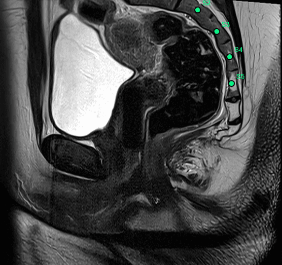
Automatic lumbosacral vertebra localization in variable field-of-view pelvic MRI
4.21 mm
Mean localization error
S1-S5
Individual sacral detection
1.4s
Per-volume inference
Multi-Center
MRI protocol robustness
Problem Statement
Automatic vertebra localization in pelvic MRI is challenging due to variable field-of-view coverage, anatomical diversity, and partial visibility of the lumbosacral spine. Manual landmark annotation is time-consuming and subject to inter-observer variability, limiting reproducibility in clinical workflows.
Approach
Developed a dual-head 3D U-Net for volumetric vertebra landmark detection in sagittal T2-weighted pelvic MRI. The system jointly predicts vertebral center heatmaps and an anatomical S1 reference point, enabling robust S1-anchored labeling across incomplete or variable spine coverage.
Results
Achieved a mean vertebral localization error of 4.21 mm across multi-center pelvic MRI datasets, with robust detection of lumbar and sacral vertebrae (L1-L5, S1-S5).
Model output · live inference
Thesis II · 17 September 2025 - 17 March 2026
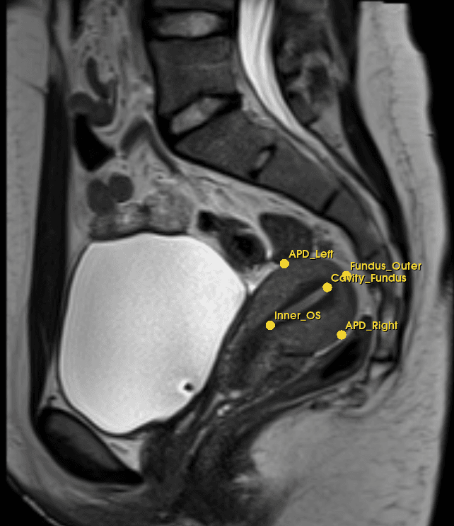
Automatic detection of uterine anatomical landmarks in pelvic MRI
4.52 mm
Overall mean error
192 landmarks evaluated
2.90 mm
Best landmark precision
Cavity Cervix
3.66 mm
Median localization error
All 6 landmarks
32
Multi-protocol test cases
3 acquisition protocols
Problem Statement
Uterine biometry — measuring fundal thickness, body length, cervical length, and AP diameter — is essential for managing endometrial cancer, fibroids, and endometriosis, yet is performed manually with high inter-observer variability. No prior automated 3D approach existed for multi-landmark localization.
Approach
Built the preprocessing pipeline used across the entire project, handling raw MRI data from three different scanner protocols. Using an nnU-Net v2 uterine segmentation as a region of interest, trained a multi-decoder 3D U-Net with a shared encoder and independent decoder branches for each landmark group, predicting six uterine landmarks simultaneously. Introduced SharpHeatmapLoss — a custom loss combining MSE with a cubic penalty term that forces sharply peaked heatmaps — and ROI-guided inference using uterine segmentation masks to eliminate background noise.
Results
Achieved 4.52 mm overall mean error across 192 landmarks and 32 test cases from 3 acquisition protocols. Cavity Cervix and Cavity Fundus reached sub-3 mm precision. Landmark detection contributed directly to a real-time reporting pipeline achieving end-to-end results in under 60 seconds, submitted to IEEE Transactions on Medical Imaging.
Model output · real predictions
Career
Work Experience
Research Assistant (Master's Thesis)
Smart Imaging Lab, Universitatsklinikum Erlangen
09/2025 - 03/2026
Erlangen, Germany
- Designed a 3D dual-decoder U-Net for automatic lumbosacral vertebral landmark localization in pelvic MRI, achieving 4.21 mm mean localization error across multi-center datasets.
- Developed dual- and triple-decoder 3D U-Net architectures for uterine landmark detection, achieving 4.52 mm mean landmark error across three MRI acquisition protocols.
- Validated deep learning models on multi-center, multi-vendor pelvic MRI datasets (0.55T-3T) across heterogeneous clinical imaging protocols.
AI Engineer
Sofitsians
04/2024 - 02/2025
Remote
- Developed AI-powered applications using LLMs, OpenAI APIs, and React.js for intelligent user experiences.
- Built and integrated RESTful APIs, RAG pipelines, and semantic search systems for AI-driven solutions.
- Implemented AI workflow automation with prompt engineering, model integration, and frontend components to streamline user interactions.
Software Developer (.NET)
Sofitsians
11/2019 - 01/2021
Islamabad, Pakistan - On-site
- Developed and maintained full-stack web applications using C#, ASP.NET MVC, and SQL Server for enterprise-level business workflows.
- Designed and optimized relational database schemas, stored procedures, and complex SQL queries to improve application performance and data reliability.
- Built and integrated REST APIs and backend services to support scalable frontend and third-party system integrations.
Research output
Publications
Selected work
Featured Projects
Technical stack
Skills
Deep Learning & AI
Programming & Deployment
AI Systems & APIs
Applied AI Engineering
Contact
Let's Connect
Have an AI engineering opportunity, research collaboration, or product idea in mind? Send a short message and I'll get back to you.